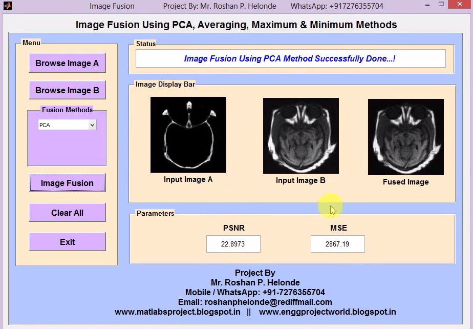
Matlab Code for Melanoma Detection / Skin Cancer Detection Using Image Processing IEEE Project

ABSTRACT
         Skin cancer – also known as malignant melanoma – is one of the deadliest form of cancer if not recognized in time. Since the pigmented areas/moles of the skin can be nicely observed by simple, non-invasive visual inspection (e.g. by a dermatoscope), the clinical protocols of its recognition also consider several visual features. Melanoma is the deadliest form of skin cancer, which is considered one of the most common human malignancies in the world. Early detection of this disease can affect the result of the illness and improve the chance of surviving. The tremendous improvement of deep learning algorithms in image recognition tasks promises a great success for medical image analysis, in particular, melanoma classification for skin cancer diagnosis. Activation functions play an important role in the performance of deep neural networks for image recognition problems as well as medical image classification. Melanin is the pigment that discerns the color of human skin. The special cells produce melanin in the skin. If these cells are damaged or unhealthy, skin discoloration is visible. Skin pigment discoloration on cheeks is a hazardous fact as a symptom of human skin disease with a possibility of losing natural beauty. The extracted information of the skin discoloration can work as a guide to diagnosis the disease. In this research, different imaging techniques like watershed method, edge detection and morphological operations are used to analyze and extract the information of cheek’s discoloration lesion by measuring the pixel number of lesion on skin. The image analyzing results are visually examined by the skin specialist and are observed to be highly accurate. The visual results are presented in the description and the accuracy of mathematical analysis is 94.88 percent.

PROJECT OUTPUT

PROJECT VIDEO

Contact:
Mr. Roshan P. Helonde
Mobile: +91-7276355704
WhatsApp: +91-7276355704
Email: roshanphelonde@rediffmail.com
Share:

Matlab Code for Currency Recognition Using Image Processing IEEE Project

ABSTRACT
           The appearance of the currency is part of this development and it is affected directly, where there is exploited in incorrect form by copying the currency in a manner similar to the reality. Therefore, it became necessary to implement a proposal for being a suitable as solution not inconsistent with the different cultures, time and place. This clear through add the watermarks inside currency, which is difficult to be copied. At the same time, this watermarks may be visible to the naked eye so can easily inferred or it is invisible. However the high resolution imaging devices can copy these additions. In this research, we have proposed a system to distinguish the currencies by the program that working a submission inferred to the watermark by feature extraction determined the type of currency. In addition to, it determined category of the currency. Benefit of it, is reducing as much as possible the spread of counterfeit currency and this system can be used by any user wants to make sure of the currency.

PROJECT OUTPUT

PROJECT VIDEO

Contact:
Mr. Roshan P. Helonde
Mobile: +91-7276355704
WhatsApp: +91-7276355704
Email: roshanphelonde@rediffmail.com
Share:

Matlab Code for Lung Cancer Detection Using Image Processing IEEE Project

ABSTRACT
        Lung cancer prevalence is one of the highest of cancers, at 18 %. One of the first steps in lung cancer diagnosis is sampling of lung tissues or biopsy. These tissue samples are then microscopically analyzed. This procedure is taken once imaging tests indicate the presence of cancer cells in the chest. Lung cancer diagnosis using lung images. One of them is that doctor still relies on subjective visual observation. A medical specialist must do thorough observation and accurate analysis in detecting lung cancer in patients. Hence, there is need for a system that is capable for detecting lung cancer automatically from microscopic images of biopsy. This method will improve the accuracy and efficiency for lung cancer detection. The aim of this research is to design a lung cancer detection system based on analysis of microscopic image of biopsy using digital image processing. Microscopic images of biopsy are feature extracted with the Gray Level Co-Occurrence Matrix (GLCM) method and classified using back propagation neural network. This method is implemented to detection of lung cancer of lung samples.

PROJECT OUTPUT

PROJECT VIDEO

Contact:
Mr. Roshan P. Helonde
Mobile: +91-7276355704
WhatsApp: +91-7276355704
Email: roshanphelonde@rediffmail.com
Share:

Matlab Code for Cheque Number Recognition Using Image Processing

ABSTRACT
          Now a days CTS bank cheques are commonly used for many financial transactions globally and processed by clearance branches by using the distinct cheque number of the corresponding bank. The proposed work focuses on segmentation and recognition of bank cheque number using optical character recognition and statistical correlation function. In general bank cheques contain the cheque number at left bottom region which will be considered as the potential area for locating and segmenting the cheque number. A standard template of numerical digits will be created from 0 to 9 for matching with the test samples using standard statistical correlation function. The manual database will be created of bank cheque images including bank cheques are used for number recognition. The accuracy of the system is analyzed by the variation on the range of the cheque image with cheque number having different font, size, quotes and varieties of characters with noise. The efficiency of the system is evaluated through the experimental results extensively. 

PROJECT OUTPUT

PROJECT VIDEO

Contact:
Mr. Roshan P. Helonde
Mobile: +91-7276355704
WhatsApp: +91-7276355704
Email: roshanphelonde@rediffmail.com
Share:

Matlab Code for Blood Group Detection Using Image Processing

ABSTRACT
          Determining of blood types is very important during emergency situation before administering a blood transfusion. Presently, these tests are performed manually by technicians, which can lead to human errors. Determination of the blood types in a short period of time and without human errors is very much essential. A method is developed based on processing of images acquired during the slide test. The image processing techniques such as thresholding and morphological operations are used. The images of the slide test are obtained from the pathological laboratory are processed and the occurrence of agglutination are evaluated. Thus the developed automated method determines the blood type using image processing techniques. The developed method is useful in emergency situation to determine the blood group without human error. The slide test consists of the mixture of one drop of blood and one drop of reagent, being the result interpreted according to the occurrence or not of agglutination. The combination of the occurrence and non occurrence of the agglutination determines the blood type of the patient. 

PROJECT OUTPUT

PROJECT VIDEO

Contact:
Mr. Roshan P. Helonde
Mobile: +91-7276355704
WhatsApp: +91-7276355704
Email: roshanphelonde@rediffmail.com
Share:

Video Steganography Matlab Project with Source Code

ABSTRACT
            Information security has become the area of concern as a result of widespread use of communication medium over the internet. This paper focuses on the data security approach when combined with encryption and steganographic techniques for secret communication by hiding it inside the multimedia files. The high results are achieved by providing the security to data before transmitting it over the internet. The files such as images, audio, video contains collection of bits that can be further translated into images, audio and video. The files composed of insignificant bits or unused areas which can be used for overwriting of other data. This Project explains the proposed algorithm using video steganography for enhancing data security. The Steganography, Cryptography and Digital Watermarking techniques can be used to obtain security and privacy of data. The steganography is the art of hiding data inside another data such as cover medium by applying different steganographic techniques. While cryptography results in making the data human unreadable form called as cipher thus cryptography is scrambling of messages. Whereas the steganography results in exploitation of human awareness so it remains unobserved and undetected or intact. It is possible to use all file medium, digital data, or files as a cover medium in steganography. Generally steganography technique is applied where the cryptography is ineffective.

PROJECT OUTPUT

PROJECT VIDEO

Contact:
Mr. Roshan P. Helonde
Mobile: +91-7276355704
WhatsApp: +91-7276355704
Email: roshanphelonde@rediffmail.com
Share:

Diabetic Retinopathy Detection Matlab Project with Source Code

ABSTRACT
                  Diabetes is a group of metabolic disease in which a person has high blood sugar.  Diabetic Retinopathy (DR) is caused by the abnormalities in the retina due to insufficient insulin in the body. It can lead to sudden vision loss due to delayed detection of retinopathy. So that Diabetic patients require regular medical checkup for effective timing of sight saving treatment.  This is continuous and stimulating research area for automated analysis of Diabetic Retinopathy in Diabetic patients. A completely automated screening system for the detection of Diabetic Retinopathy can effectively reduces the burden of the specialist and saves cost as well as time. Due to noise and other disturbances that occur during image acquisition Diabetic Retinopathy may lead to false detection and this is overcome by various image processing techniques. Further the different features are extracted which serves as the guideline to identify and grade the severity of the disease. Based on the extracted features classification of the retinal image as normal or abnormal is carried out.

PROJECT OUTPUT

PROJECT VIDEO

Contact:
Mr. Roshan P. Helonde
Mobile: +91-7276355704
WhatsApp: +91-7276355704
Email: roshanphelonde@rediffmail.com
Share:

Image Steganography Using Matlab Project with Source Code

ABSTRACT
          Steganography is the one type of powerful technique which is science & art in which we have to write hidden messages, or we hide some important images, audio files, videos in this way that no-one, can find a hidden message which exists in cover images. Steganography is most strong techniques to mask the existence of unseen secret data within a cover object. Actually Stego means "Cover" graphy means "writing" that means It is nothing but we are hiding secret objects in cover image in which medium is different types of images. In practical feasible implementation practical approach would be to make the algorithm as strong as possible. In steganographed images are the most powerful objects that means cover objects, and therefore importance of image steganographed which can Embedding secret information inside images requires systematic computations. Various metrics were used to judge imperceptibility of steganography. The metrics in Matlab indicates how similar or dissimilar the stego-image compares with Cover.

PROJECT OUTPUT

PROJECT VIDEO

Contact:
Mr. Roshan P. Helonde
Mobile: +91-7276355704
WhatsApp: +91-7276355704
Email: roshanphelonde@rediffmail.com
Share:

Real Time Face Recognition Using Matlab Project with Source Code

ABSTRACT
             The subject of face recognition is as old as computer vision because of the practical importance of the topic and theoretical interest from cognitive scientists. Despite the fact that other methods of identification (such as fingerprints, or iris scans) can be more accurate, face recognition has always remains a major focus of research because of its noninvasive nature and because it is people's primary method of person identification. This electronic document is about face detection. In computer literature face detection has been one of the most studied topics. Given an arbitrary image, the goal of this project is to determine real time face recognition. While this appears to be a trivial task for human beings, it is very challenging task for computers. The difficulty associated with face detection can be attributed to many variations in scale, location, view point, illumination, occlusions, etc. Although there have been hundreds of reports reported approaches for face detection, if one were asked to name a single face detection algorithm that has most impact in recent decades, it will most likely be the face detection, which is capable of processing images extremely rapidly and achieve high detection rates.

PROJECT VIDEO

PROJECT VIDEO

Contact:
Mr. Roshan P. Helonde
Mobile: +91-7276355704
WhatsApp: +91-7276355704
Email: roshanphelonde@rediffmail.com
Share:

Fruit Disease Detection and Classification Using Image Processing Matlab Project with Source Code

ABSTRACT
            Diseases in fruit cause devastating problem in economic losses and production in agricultural industry worldwide. In this paper, a solution for the detection and classification of fruit diseases is proposed and experimentally validated. The image processing based proposed approach is composed of the following steps; in the first step K-Means clustering technique is used for the image segmentation, in the second step some features are extracted from the segmented image, and finally images are classified into one of the classes by using a Support Vector Machine. Our experimental results express that the proposed solution can significantly support accurate detection and automatic classification of fruit diseases.
             Fruit diseases can cause significant losses in yield and quality appeared in harvesting. For example, soybean rust (a fungal disease in soybeans) has caused a significant economic loss and just by removing 20% of the infection, the farmers may benefit with an approximately 11 million-dollar profit (Roberts et al., 2006). Some fruit diseases also infect other areas of the tree causing diseases of twigs, leaves and branches. An early detection of fruit diseases can aid in decreasing such losses and can stop further spread of diseases. A lot of work has been done to automate the visual inspection of the fruits by machine vision with respect to size and color. However, detection of defects in the fruits using images is still problematic due to the natural variability of skin color in different types of fruits, high variance of defect types, and presence of stem/calyx. To know what control factors to consider next year to overcome similar losses, it is of great significance to analyze what is being observed.

PROJECT OUTPUT

PROJECT VIDEO

Contact:
Mr. Roshan P. Helonde
Mobile: +91-7276355704
WhatsApp: +91-7276355704
Email: roshanphelonde@rediffmail.com
Share:

Real Time Driver Drowsiness Detection Using Matlab Project with Source Code

ABSTRACT
             Drivers who do not take regular breaks when driving long distances run a high risk of becoming drowsy and cause accidents. It is a state which they often fail to recognise early enough according to the experts. Studies show that around one quarter of all serious motorway accidents is attributable to sleepy drivers in need of a rest, meaning that drowsiness causes more road accidents than drink-driving. Driver fatigue is a significant factor in a large number of vehicle accidents. The development of technologies for detecting drowsiness at the wheel is a major challenge in the field of accident avoidance systems. Because of the hazard that drowsiness presents on the road, methods need to be developed for counteracting its affects. The main aim of this is to develop a drowsiness detection system by monitoring the eyes and mouth; it is believed that the symptoms of driver fatigue can be detected early enough to avoid a car accident. Detection of fatigue involves the observation of eye movements, blink patterns and mouth opening for yawning. The analysis of face images is a popular research area with applications such as face recognition, and human identification security systems. This project is focused on the localization of the eyes, which involves looking at the entire image of the eye, and determining the position of the eyes.

PROJECT OUTPUT

PROJECT VIDEO

Contact:
Mr. Roshan P. Helonde
Mobile: +91-7276355704
WhatsApp: +91-7276355704
Email: roshanphelonde@rediffmail.com
Share:

Brain Tumor Detection and Classification Using Neural Network Matlab Project with Source Code

ABSTRACT
          The imaging plays a central role in the diagnosis of brain tumors. An efficient algorithm is proposed in this project for brain tumor detection based on digital image segmentation. Brain tumor may be considered among the most difficult tumors to treat, as it involves the organ which is not only in control of the body. We proposed an Artificial Neural Network Approach for Brain Tumor Detection, which gave the edge pattern and segment of brain and brain tumor itself. The segmentation of brain tumors in magnetic resonance images is a challenging and difficult task because of the variety of their possible shapes, locations, image intensities. In this project it is intended to summarize and compare the methods of automatic detection of brain tumor through Magnetic Resonance Image used in different stages of Computer Aided Detection System. Brain Image classification techniques are studied. Existing methods are classically divided into region based and contour based methods. These are usually dedicated to full enhanced tumors or specific types of tumors. The amount of resources required to describe large set of data is simplified and selected in for tissue segmentation. In this project image segmentation techniques were applied on input images in order to detect brain tumors. Also in this project a Neural Network model that is based on machine learning with image and data analysis and manipulation techniques is proposed to carry out an automated brain tumor classification.

PROJECT OUTPUT

PROJECT VIDEO

Contact:
Mr. Roshan P. Helonde
Mobile: +91-7276355704
WhatsApp: +91-7276355704
Email: roshanphelonde@rediffmail.com
Share:

Diabetic Retinopathy Detection Using Matlab Code

ABSTRACT
                  Diabetes is a group of metabolic disease in which a person has high blood sugar.  Diabetic Retinopathy (DR) is caused by the abnormalities in the retina due to insufficient insulin in the body. It can lead to sudden vision loss due to delayed detection of retinopathy. So that Diabetic patients require regular medical checkup for effective timing of sight saving treatment.  This is continuous and stimulating research area for automated analysis of Diabetic Retinopathy in Diabetic patients. A completely automated screening system for the detection of Diabetic Retinopathy can effectively reduces the burden of the specialist and saves cost as well as time. Due to noise and other disturbances that occur during image acquisition Diabetic Retinopathy may lead to false detection and this is overcome by various image processing techniques. Further the different features are extracted which serves as the guideline to identify and grade the severity of the disease. Based on the extracted features classification of the retinal image as normal or abnormal is carried out.  In this paper, we have presented detail study of various screening methods for Diabetic Retinopathy. Many researchers have made number of attempts to improve accuracy, productivity, sensitivity and specificity.

PROJECT OUTPUT

PROJECT VIDEO


Contact:
Mr. Roshan P. Helonde
Mobile: +91-7276355704
WhatsApp: +91-7276355704
Email: roshanphelonde@rediffmail.com
Share:

Iris Recognition Using Image Processing Matlab Code IEEE Based Project

ABSTRACT
               This project presents an iris coding method for effective recognition of an individual. The recognition is performed based on a mathematical and computational method called discrete cosine transform (DCT). It consists of calculating the differences of discrete cosine transform (DCT) coefficients of overlapped angular patches from the normalized iris image for the purpose of feature extraction. DCT is used because it offers efficiency, it is much more practical and its basis vectors are comprised of entirely real-valued components. Iris recognition belongs to the biometric identification. Biometric identification is a technology that is used for the identification an individual based on ones physiological or behavioral characteristics. Iris is the strongest physiological feature for the recognition process because it offers most accurate and reliable results. Iris recognition process mainly involves three stages namely, iris image preprocessing, feature extraction and template matching. In the pre-processing step, iris localization algorithm is used to locate the inner and outer boundaries of the iris. Detected iris region is then normalized to a fixed size rectangular block. In the feature extraction step, texture analysis method is used to extract significant features from the normalized iris image with the help of Discrete Cosine Transform (DCT).

PROJECT OUTPUT

PROJECT VIDEO


Contact:
Mr. Roshan P. Helonde
Mobile: +91-7276355704
WhatsApp: +91-7276355704
Email: roshanphelonde@rediffmail.com
Share:

Character Recognition from Images Using Matlab Code IEEE Based Project

ABSTRACT
                 Optical character recognition (OCR) is becoming a powerful tool in the field of Character Recognition, now a days. In the existing globalized environment, OCR can play a vital role in different application fields. Basically, OCR technique converts images into editable format. This technique converts images in the form of documents such as we can edit, modify and store data more safely for longtime. This paper presents basic of OCR technique with its components such as pre-processing, Feature Extraction, Classification, post-processing etc. There are various techniques have been implemented for the recognition of character. This Review also discusses different ideas implemented earlier for recognition of a character. This project may act as a supportive material for those who wish to know about OCR. Now a days, globalization is reaching to a great level. In this globalized environment, character recognition techniques also getting a valuable demand in number of application areas. OCR is an effective technique which converts image into suitable format such that data can be edit, modify and stored. This technique performs several operations such as, scans the input image, processes over the scanned image thereby image gets converted into portable formats .For instance, the hard copy of old historical books, novels, etc. .cannot be stored safely for a long time. Rather, its safety has limitations. If we apply OCR technique for such cases, the different historical documents can be stored, modified for a longtime. OCR also having variety of applications in almost all fields, including security. OCR implementation helps us to edit, store and process over the scanned data more effectively. User can handle the stored data whenever he wants with the internet support. So Optical character recognition is most successful application used in pattern recognition.

PROJECT OUTPUT

PROJECT VIDEO


Contact:
Mr. Roshan P. Helonde
Mobile: +91-7276355704
WhatsApp: +91-7276355704
Email: roshanphelonde@rediffmail.com
Share:

Brain Tumor Detection on MRI Images Using Segmentation and Clustering Full Matlab Project Code

ABSTRACT
          Image processing is an active research area in which medical image processing is a highly challenging field. Medical imaging techniques are used to image the inner portions of the human body for medical diagnosis. Brain tumor is a serious life altering disease condition. Image segmentation plays a significant role in image processing as it helps in the extraction of suspicious regions from the medical images. In this paper we have proposed segmentation of brain MRI image using K-means clustering algorithm followed by morphological filtering which avoids the mis-clustered regions that can inevitably be formed after segmentation of the brain MRI image for detection of tumor location. In this paper, we present a system based on gabor filter based enhancement technique and feature extraction techniques using texture based segmentation and SOM (Self Organization Map) which is a form of Artificial Neural Network (ANN) used to analyze the texture features extracted. SOM determines which texture feature has the ability to classify benign, malignant and normal cases. Watershed segmentation technique is used to classify cancerous region from the non cancerous region.

PROJECT OUTPUT

PROJECT VIDEO


Contact:
Mr. Roshan P. Helonde
Mobile: +91-7276355704
WhatsApp: +91-7276355704
Email: roshanphelonde@rediffmail.com
Share:

Signature Recognition and Verification Using Matlab Project Code

ABSTRACT
         The fact that the signature is widely used as a means of personal identification tool for humans require that the need for an automatic verification system. Verification can be performed either Offline or Online based on the application. However human signatures can be handled as an image and recognized using computer vision and neural network techniques. With modern computers, there is need to develop fast algorithms for signature recognition. There are various approaches to signature recognition with a lot of scope of research. Signature verification and recognition is a technology that can improve security in our day to day transaction held in society. This project presents a novel approach for offline signature verification. In this project signature verification using Image Processing is projected, where the signature is written on a paper are obtained using a scanner or a camera captured and presented in an image format. For authentication of signature, the proposed method is based on geometrical and statistical feature extraction and then the entire database, features are trained using neural network. The extracted features of investigation signature are compared with the previously trained features of the reference signature. This technique is suitable for various applications such as bank transactions, passports with good authentication results etc.

PROJECT OUTPUT

PROJECT VIDEO


Contact:
Mr. Roshan P. Helonde
Mobile: +91-7276355704
WhatsApp: +91-7276355704
Email: roshanphelonde@rediffmail.com
Share:

Audio Compression Using Matlab Project Code

ABSTRACT
         Speech compression is the technique of converting human speech into an efficiently encoded format that can later be decoded to produce a close approximation of the original signal. The merits of the compression technique are reduction in storage space, bandwidth, transmission power and energy. An efficient algorithm Discrete Wavelet Transform (DWT) is employed for decomposition of original signal into wavelets coefficients at different scales and positions and these coefficients are truncated to perform encoding and decoding. The compression technique used in this project is better than other earlier coding techniques like μ-law coding, code excited linear predictive coding. Speech compression plays a prominent role in speech signal processing such as satellite communications, internet communications, transmission of biomedical signals and other applications. Wavelet is one of the recent developments to overcome the limitations of Fourier transform of signal analysis which has the special ability to examine signal simultaneously in both time and frequency. Wavelet analysis is breaking up of an original signal into scaled and shifted versions called the mother wavelet. Wavelet Transform has emerged as a recent powerful mathematical tool in many areas of science and technology, more so in the field of signal processing.

PROJECT OUTPUT

PROJECT VIDEO


Contact:
Mr. Roshan P. Helonde
Mobile: +91-7276355704
WhatsApp: +91-7276355704
Email: roshanphelonde@rediffmail.com
Share:

Cotton Leaf Disease Detection and Classification Using Matlab Code

ABSTRACT
            Cotton is one of the most important fiber crop which is used as raw material in textile industries. But, now-a-days cotton is facing number of problems related to the healthy growth of crop due to diseases. These diseases are reducing the productivity of cotton crop and farmers are getting suffered financially due to this crop loss. Agriculture is an important source of livelihood where 65% population is depend on it. The crop loss due to disease is increasing day by day which affects on the quality and productivity of crop. As diseases on the crop are certain, the early disease detection of the crop plays major role to control the loss in agriculture. In the proposed disease detection system, the work is carried out on cotton leaves. Initially the infected region is captured and pre-processed. During segmentation, leaf as well as diseased part is segmented using k means clustering method and different features are extracted such as color and texture with the help of color-co-occurrence method. Finally classification technique is used for detecting the diseases with the help of SVM (Support Vector Machine) classifier.

PROJECT OUTPUT

PROJECT VIDEO


Contact:
Mr. Roshan P. Helonde
Mobile: +91-7276355704
WhatsApp: +91-7276355704
Email: roshanphelonde@rediffmail.com
Share:

Brain Tumor Detection on MRI Images Using Matlab Project Code

ABSTRACT
          Image processing is an active research area in which medical image processing is a highly challenging field. Medical imaging techniques are used to image the inner portions of the human body for medical diagnosis. Brain tumor is a serious life altering disease condition. Image segmentation plays a significant role in image processing as it helps in the extraction of suspicious regions from the medical images. In this project we have proposed segmentation of brain MRI image using K-means clustering algorithm followed by morphological filtering which avoids the mis-clustered regions that can inevitably be formed after segmentation of the brain MRI image for detection of tumor location. In this project, we present a system based on enhancement technique and feature extraction techniques using segmentation and clustering used to analyze the texture features extracted texture feature has the ability to classify benign, malignant.

PROJECT OUTPUT

PROJECT VIDEO


Contact:
Mr. Roshan P. Helonde
Mobile: +91-7276355704
WhatsApp: +91-7276355704
Email: roshanphelonde@rediffmail.com
Share:

Lung Cancer Detection Using Image Processing Matlab Project Code

ABSTRACT
              The most common cause of lung cancer is long‐term exposure to tobacco smoke, which causes 80‐90% of lung cancers. Cancer cells can be carried away from the lungs in blood, or lymph fluid that surrounds lung tissue. Lymph flows through lymphatic vessels, which drain into lymph nodes located in the lungs and in the center of the chest. Lung cancer often spreads toward the center of the chest because the natural flow of lymph out of the lungs is toward the center of the chest. One of the major reason for non-accidental death is cancer. It has been proved that lung cancer is the topmost cause of cancer death in men and women worldwide. The death rate can be reduced if people go for early diagnosis so that suitable treatment can be administered by the clinicians within specified time. Cancer is, when a group of cells go irregular growth uncontrollably and lose balance to form malignant tumors which invades surrounding tissues. Cancer can be classified as Non-small cell lung cancer and small cell lung cancer. The various ways to detect lung cancer is by the use of image processing , pattern recognition and artificial neutral network to develop Computer aided diagnosis. In this project we use the techniques and algorithm used in image processing to detect cancer in three types of medical images. In this system first of all the medical images are recorded using a suitable imaging system. The images obtained are taken as input for the system where the image first go through the various steps of image processing like pre-processing, edge detection, morphological processing ,feature extraction.
                   Lung cancer which is among the five main types of cancer is a leading one to overall cancer mortality. Cancer is a serious health problem among various kinds of diseases. World Health Organization (WHO) reports that worldwide 7.6 million deaths are caused by cancer each year. Uncontrollable cell development in the tissues of the lung is called as lung cancer. Lung nodule is an abnormality that leads to lung cancer, characterized by a small round or oval shaped growth on the lung which appears as a white shadow in the CT scan. These uncontrollable cells restrict the growth of healthy lung tissues. If not treated, this growth can spread beyond the lung in the nearby tissue called metastasis and, form tumors. In order to preserve the life of the people who are suffered by the lung cancer disease, it should be pre‐diagnosed. The overall 5‐year survival rate for lung cancer patients increases from 14 to 49% if the disease is detected in time. So there is a need of pre-diagnosis system for lung cancer disease which should provide better results.

PROJECT OUTPUT

PROJECT VIDEO


Contact:
Mr. Roshan P. Helonde
Mobile: +91-7276355704
WhatsApp: +91-7276355704
Email: roshanphelonde@rediffmail.com
Share:

Anemia Diseases Detection Present in Blood Cells Smear Using Matlab Project

ABSTRACT
          Anemia is a blood disorder which results from the abnormalities of red blood cells and shortens the life expectancy to 42 and 48 years for males and females respectively. It also causes pain jaundice, shortness of breath, etc. Anemia is characterized by the presence of abnormal cells like sickle cell, ovalocyte, anisopoikilocyte. Sickle cell disease usually presenting in childhood, occurs more commonly in people from parts of tropical and subtropical regions where malaria is or was very common. A healthy RBC is usually round in shape. But sometimes it changes its shape to form a sickle cell structure; this is called as sickling of RBC. Majority of the sickle cells (whose shape is like crescent moon) found are due to low haemoglobin content. An image processing algorithm to automate the diagnosis of sickle-cells present in thin blood smears is developed. Images are acquired using a charge-coupled device camera connected to a light microscope. Clustering based segmentation techniques are used to identify erythrocytes (red blood cells) and Sickle-cells present on microscopic slides. Image features based on colour, texture and the geometry of the cells are generated, as well as features that make use of a priori knowledge of the classification problem and mimic features used by human technicians. The proposed image processing based identification of sickle-cells in anemic patient will be very helpful for automatic, sleek and effective diagnosis of the disease.

PROJECT OUTPUT

PROJECT VIDEO


Contact:
Mr. Roshan P. Helonde
Mobile: +91-7276355704
WhatsApp: +91-7276355704
Email: roshanphelonde@rediffmail.com

Share:

Emotion Recognition from Audio Signal Full Matlab Project Code

ABSTRACT
          Emotion Recognition is a recent research topic in the field of Human Computer Interaction Intelligence and mostly used to develop wide range of applications such as stress management for call centre employee, and learning & gaming software, In E-learning field, identifying students emotion timely and making appropriate treatment can enhance the quality of teaching. Main aim of HCI is to achieve a more natural interaction between machine and humans. HCI is an emerging field using which we can improve the interactions between users and computers by making computers more respond able to the user’s needs. Today’s HCI system has been developed to identify who is speaking or what he/she is speaking. If in the HCI system, the computers are given an ability to detect human emotions then they can know how he/she is speaking and can respond accurately and naturally like humans do. In this project methodology for emotion recognition from speech signal is presented. Here, some of acoustic features are extracted from speech signal to analyze the characteristics and behavior of speech. The system is used to recognize the basic emotions. It can serve as a basis for further designing an application for human like interaction with machines through natural language processing and improving the efficiency of emotion. In this format, energy, Mel Frequency Cepstral Coefficients (MFCC) has been used for feature extraction from the speech signal. Support Vector Machine (SVM) are used for recognition of emotional states. English datasets are used for analysis of emotions with SVM Kernel functions.

PROJECT OUTPUT

PROJECT VIDEO


Contact:
Mr. Roshan P. Helonde
Mobile: +91-7276355704
WhatsApp: +91-7276355704
Email: roshanphelonde@rediffmail.com
Share:

Image Fusion Using PCA on MRI and CT Image Full Matlab Project Code

ABSTRACT
           Image Fusion is a procedure of merging the required image information from two or more images of the same scene or background into a single picture and the resultant fused picture will be more informational and complete than any of the input images, which was taken for fusion. Processing image could be multi sensor, multi-modal, multi-focal or multi passing. Image fusion is a strategy that combine complimentary details from two or more input image such that the new image gives more information and more suitable for the motivation behind human visual system. This project presents a survey on a percentage of the image fusion technique (simple average, simple minimum, simple maxima, PCA, DWT). Comparative examination of the considerable number of systems closes the better approach for its future exploration.

PROJECT OUTPUT

PROJECT VIDEO


Contact:
Mr. Roshan P. Helonde
Mobile: +91-7276355704
WhatsApp: +91-7276355704
Email: roshanphelonde@rediffmail.com
Share:

Melanoma Detection / Skin Cancer Detection Using Image Processing Full Matlab Project Code

ABSTRACT
         Skin cancer – also known as malignant melanoma – is one of the deadliest form of cancer if not recognized in time. Since the pigmented areas/moles of the skin can be nicely observed by simple, non-invasive visual inspection (e.g. by a dermatoscope), the clinical protocols of its recognition also consider several visual features. Melanoma is the deadliest form of skin cancer, which is considered one of the most common human malignancies in the world. Early detection of this disease can affect the result of the illness and improve the chance of surviving. The tremendous improvement of deep learning algorithms in image recognition tasks promises a great success for medical image analysis, in particular, melanoma classification for skin cancer diagnosis. Activation functions play an important role in the performance of deep neural networks for image recognition problems as well as medical image classification. Melanin is the pigment that discerns the color of human skin. The special cells produce melanin in the skin. If these cells are damaged or unhealthy, skin discoloration is visible. Skin pigment discoloration on cheeks is a hazardous fact as a symptom of human skin disease with a possibility of losing natural beauty. The extracted information of the skin discoloration can work as a guide to diagnosis the disease. In this research, different imaging techniques like watershed method, edge detection and morphological operations are used to analyze and extract the information of cheek’s discoloration lesion by measuring the pixel number of lesion on skin. The image analyzing results are visually examined by the skin specialist and are observed to be highly accurate. The visual results are presented in the description and the accuracy of mathematical analysis is 94.88 percent.

PROJECT OUTPUT

PROJECT VIDEO


Contact:
Mr. Roshan P. Helonde
Mobile: +91-7276355704
WhatsApp: +91-7276355704
Email: roshanphelonde@rediffmail.com
Share:

Brain Tumor Detection and Classification Using Matlab Project Code

ABSTRACT
          Brain tumors are the most common issue in children. Approximately 3,410 children and adolescents under age 20 are diagnosed with primary brain tumors each year. Brain tumors, either malignant or benign, that originate in the cells of the brain. The conventional method of detection and classification of brain tumor is by human inspection with the use of medical resonant brain images. But it is impractical when large amounts of data is to be diagnosed and to be reproducible. And also the operator assisted classification leads to false predictions and may also lead to false diagnose. Medical Resonance images contain a noise caused by operator performance which can lead to serious inaccuracies classification. The use Support Vector Machine, Kmean and PCA shown great potential in this field. Principal Component Analysis gives fast and accurate tool for Feature Extraction of the tumors. Kmean is used for Image Segmentation. Support Vector Mahine with Training and Testing of Brain Tumor Images techniques is implemented for automated brain tumor classification.

PROJECT OUTPUT

PROJECT VIDEO


Contact:
Mr. Roshan P. Helonde
Mobile: +91-7276355704
WhatsApp: +91-7276355704
Email: roshanphelonde@rediffmail.com
Share:

Audio Steganography Using Matlab Project Code

ABSTRACT
            Steganography is the art and science of writing hidden messages in such a way that no one, apart from the sender and intended recipient, suspects the existence of the message, a form of security through obscurity. Steganography works by replacing bits of useless or unused data in regular computer files (such as graphics, sound, text, HTML, or even floppy disks ) with bits of different, invisible information. This hidden information can be plain text, cipher text, or even images. The rapid development of multimedia and internet allows for wide distribution of digital media data. It becomes much easier to edit, modify and duplicate digital information. Besides that, digital documents are also easy to copy and distribute, therefore it will be faced by many threats. It is a big security and privacy issue, it become necessary to find appropriate rotation because of the significance, accuracy and sensitivity of the information. Steganography and Cryptography are considered as one of the techniques which are used to protect the important information, but both techniques have their pro’s and con’s. In this proposed system of audio steganography we have implemented a new scheme based on mel frequency components. The mel frequency cepstrum coefficients are used for finding the unique feature audio data in audio file. The returned features provide us with highly robust and high end features with low invariance. We have used this property of MFCC in order to detect high bandwidth free space location in the sound data and have embedded the encrypted watermark image data into these MFCC components. The proposed scheme works to increase the PSNR values and reduce the error rate of hiding the data in the image and thus improves the sound quality and makes it look original. The effect of MFCC is positive as the watermark extracted from this proposed scheme shows high correlation to the original watermark and also the resistance towards various attacks has also been improved, the attacks degrade the watermark due to high payload or bigger watermark size, the probability of extraction of watermark or steganograph data becomes higher.

PROJECT OUTPUT

PROJECT VIDEO


Contact:
Mr. Roshan P. Helonde
Mobile: +91-7276355704
WhatsApp: +91-7276355704
Email: roshanphelonde@rediffmail.com
Share:

Currency Recognition Using Matlab Project Code

ABSTRACT
           The appearance of the currency is part of this development and it is affected directly, where there is exploited in incorrect form by copying the currency in a manner similar to the reality. Therefore, it became necessary to implement a proposal for being a suitable as solution not inconsistent with the different cultures, time and place. This clear through add the watermarks inside currency, which is difficult to be copied. At the same time, this watermarks may be visible to the naked eye so can easily inferred or it is invisible. However the high resolution imaging devices can copy these additions. In this research, we have proposed a system to distinguish the currencies by the program that working a submission inferred to the watermark by feature extraction determined the type of currency. In addition to, it determined category of the currency. Benefit of it, is reducing as much as possible the spread of counterfeit currency and this system can be used by any user wants to make sure of the currency.

PROJECT OUTPUT

PROJECT VIDEO


Contact:
Mr. Roshan P. Helonde
Mobile: +91-7276355704
WhatsApp: +91-7276355704
Email: roshanphelonde@rediffmail.com
Share:

Lung Cancer Detection Using Matlab Project Code

ABSTRACT
        Lung cancer prevalence is one of the highest of cancers, at 18 %. One of the first steps in lung cancer diagnosis is sampling of lung tissues or biopsy. These tissue samples are then microscopically analyzed. This procedure is taken once imaging tests indicate the presence of cancer cells in the chest. Lung cancer diagnosis using lung images. One of them is that doctor still relies on subjective visual observation. A medical specialist must do thorough observation and accurate analysis in detecting lung cancer in patients. Hence, there is need for a system that is capable for detecting lung cancer automatically from microscopic images of biopsy. This method will improve the accuracy and efficiency for lung cancer detection. The aim of this research is to design a lung cancer detection system based on analysis of microscopic image of biopsy using digital image processing. Microscopic images of biopsy are feature extracted with the Gray Level Co-Occurrence Matrix (GLCM) method and classified using back propagation neural network. This method is implemented to detection of lung cancer of lung samples.

PROJECT OUTPUT

PROJECT VIDEO


Contact:
Mr. Roshan P. Helonde
Mobile: +91-7276355704
WhatsApp: +91-7276355704
Email: roshanphelonde@rediffmail.com
Share:

Tomato Disease Detection and Classification Using Matlab Project Code

ABSTRACT
       Agriculture is the major sector in India. About 58% of the rural livelihood influenced by in agriculture. Out of which tomato is one of the common food crops in India. Due to which detection of disease on tomato plant becomes important because less susceptibility. The plants productivity gets affected if proper care is not taken. Image processing is one of upbringing technology which is helping to resolve such issues with various algorithms and techniques. Most of the diseases of tomato disease detected at initial stages. By detecting the diseases at initial stage on tomatos will surely avoid impending loss. In this project four key diseases are identified using image segmentation and Multi-class SVM algorithm. For parting of damaged area of tomato image segmentation is used and for classification of accurate disease Multi-class SVM algorithm is used. 

PROJECT OUTPUT

PROJECT VIDEO


Contact:
Mr. Roshan P. Helonde
Mobile: +91-7276355704
WhatsApp: +91-7276355704
Email: roshanphelonde@rediffmail.com
Share:

Contact Us

Name

Email *

Message *

Blog Archive

Blog Archive

Popular posts